Cell Injury: Reversible, Irreversible, and How Cells Die
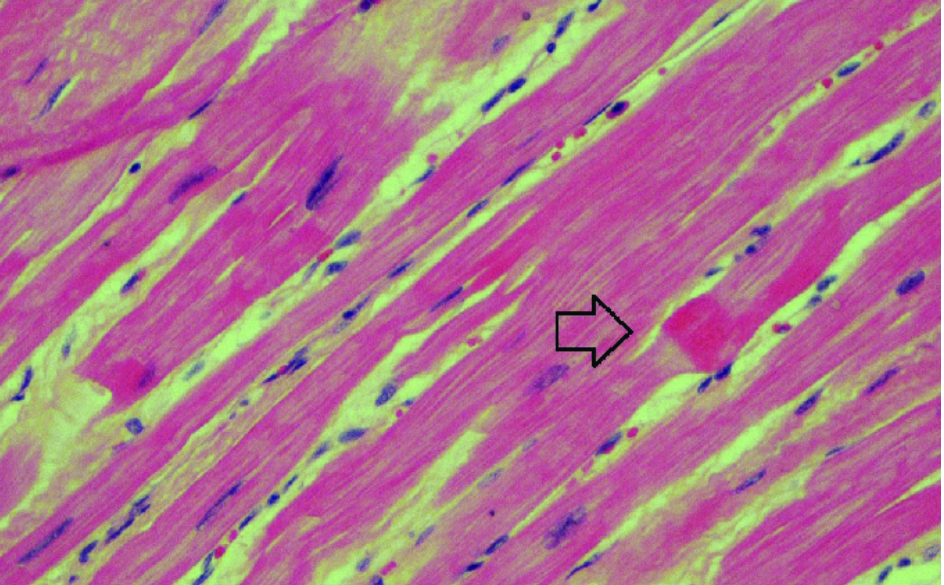
Every disease begins at the level of the cell. When a cell is pushed beyond its ability to adapt, it gets injured. If the stress is mild or short, the cell recovers. If it is severe or prolonged, the cell dies. Understanding this sequence is the foundation of general pathology.
The spectrum of cellular response
- Normal cell – in homeostasis.
- Adaptation – hypertrophy, hyperplasia, atrophy, metaplasia, when stress is sustained but tolerable.
- Reversible injury – function and structure disturbed, but recovery is possible if the cause is removed.
- Irreversible injury → cell death – necrosis or apoptosis.
The key question in any injury: has the cell crossed the point of no return?
Causes of cell injury
| Category | Examples |
|---|---|
| Oxygen deprivation | Hypoxia, ischemia, anemia, CO poisoning |
| Physical agents | Trauma, heat, cold, radiation, electric shock |
| Chemicals and drugs | Paracetamol overdose, alcohol, CCl₄, heavy metals |
| Infectious agents | Viruses, bacteria, fungi, parasites |
| Immunologic reactions | Autoimmunity, hypersensitivity |
| Genetic defects | Enzyme deficiencies, misfolded proteins |
| Nutritional imbalance | Protein–calorie deficiency, vitamin deficiency, obesity |
Hypoxia vs ischemia: ischemia (loss of blood supply) damages tissue faster than hypoxia alone, because it also cuts off glucose and allows toxic metabolites to accumulate, so even anaerobic glycolysis stops.
Reversible injury
Light microscopy
- Cellular swelling (hydropic change) – the earliest visible change. Cells look pale and enlarged because of failure of the Na⁺/K⁺ ATPase pump and water influx.
- Fatty change – small or large lipid vacuoles in the cytoplasm, seen mainly in cells involved in fat metabolism (hepatocytes, myocardium).
Electron microscopy
- Plasma membrane blebbing and loss of microvilli
- Mitochondrial swelling with small amorphous densities
- Dilated endoplasmic reticulum with detachment of ribosomes
- Nucleolar alterations, clumping of chromatin
- Myelin figures (whorls of phospholipid from damaged membranes)
Irreversible injury – the point of no return
Two events define irreversibility:
- Inability to restore mitochondrial function – large, flocculent amorphous densities appear in mitochondria.
- Profound membrane damage – of the plasma membrane and lysosomal membranes, causing leakage of cellular contents and self-digestion.
Nuclear changes of cell death
| Change | Meaning |
|---|---|
| Pyknosis | Nucleus shrinks and becomes dense and dark |
| Karyorrhexis | Pyknotic nucleus breaks into fragments |
| Karyolysis | Nuclear staining fades as DNA is digested |
The cytoplasm becomes more eosinophilic (loss of RNA, denatured proteins).
Mechanisms of cell injury
1. ATP depletion
- Na⁺/K⁺ pump fails → sodium and water enter → cell swelling
- Shift to anaerobic glycolysis → lactic acid → fall in pH → chromatin clumping
- Ribosome detachment → reduced protein synthesis
2. Mitochondrial damage
- Opening of the mitochondrial permeability transition pore → loss of membrane potential
- Leakage of cytochrome c → triggers apoptosis
3. Calcium influx Excess cytosolic calcium activates damaging enzymes:
- Phospholipases → membrane damage
- Proteases → cytoskeletal breakdown
- Endonucleases → DNA fragmentation
- ATPases → further ATP loss
4. Oxidative stress (free radicals)
- Superoxide, hydrogen peroxide, hydroxyl radical
- Cause lipid peroxidation of membranes, protein damage, DNA breaks
- Defenses: superoxide dismutase, catalase, glutathione peroxidase, vitamins E and C
5. Membrane damage – the decisive event leading to cell death.
6. DNA damage and protein misfolding – when repair fails, the cell activates apoptosis.
Types of necrosis
| Type | Where seen | Key feature |
|---|---|---|
| Coagulative | Infarcts of heart, kidney, spleen | Cell outlines preserved, nuclei lost |
| Liquefactive | Brain infarct, abscess | Tissue digested into a liquid mass |
| Caseous | Tuberculosis | Cheese-like, granular pink debris within a granuloma |
| Fat | Acute pancreatitis | Chalky white areas from saponification |
| Fibrinoid | Vessel walls in vasculitis, malignant hypertension | Bright pink, fibrin-like deposits |
| Gangrenous | Limbs, bowel (clinical term) | Coagulative ± liquefactive (wet) necrosis |
Necrosis vs apoptosis
| Feature | Necrosis | Apoptosis |
|---|---|---|
| Cell size | Enlarged (swelling) | Reduced (shrinkage) |
| Nucleus | Pyknosis, karyorrhexis, karyolysis | Condensation, fragmentation |
| Plasma membrane | Disrupted | Intact |
| Cell contents | Leak out | Packed into apoptotic bodies |
| Inflammation | Present | Absent |
| Nature | Always pathological | Physiological or pathological |
Clinical relevance
Leakage of intracellular proteins through damaged membranes is the basis of routine laboratory diagnosis:
- Cardiac troponins, CK-MB – myocardial injury
- ALT, AST – hepatocyte injury
- Amylase, lipase – pancreatic injury
In the myocardium, loss of contractility occurs within about a minute of ischemia, and injury becomes irreversible after roughly 20–30 minutes. This is why early reperfusion in myocardial infarction saves muscle.
Quick revision points
- Earliest light-microscopic change: cellular swelling
- Hallmarks of irreversibility: mitochondrial dysfunction + membrane damage
- Brain infarct shows liquefactive, not coagulative, necrosis
- Apoptosis: no inflammation, membrane intact
- Enzyme leakage in blood = evidence of irreversible injury
Test yourself
The earliest change seen under the light microscope in reversible cell injury is:
Which feature best indicates irreversible cell injury?
Infarction of the brain typically shows which type of necrosis?
Fat necrosis with saponification is characteristically seen in:
Which feature favours apoptosis over necrosis?
Questions or corrections are welcome. This post is for learning and teaching; always refer to standard textbooks for clinical decisions.