Autophagy: How Cells Recycle Themselves
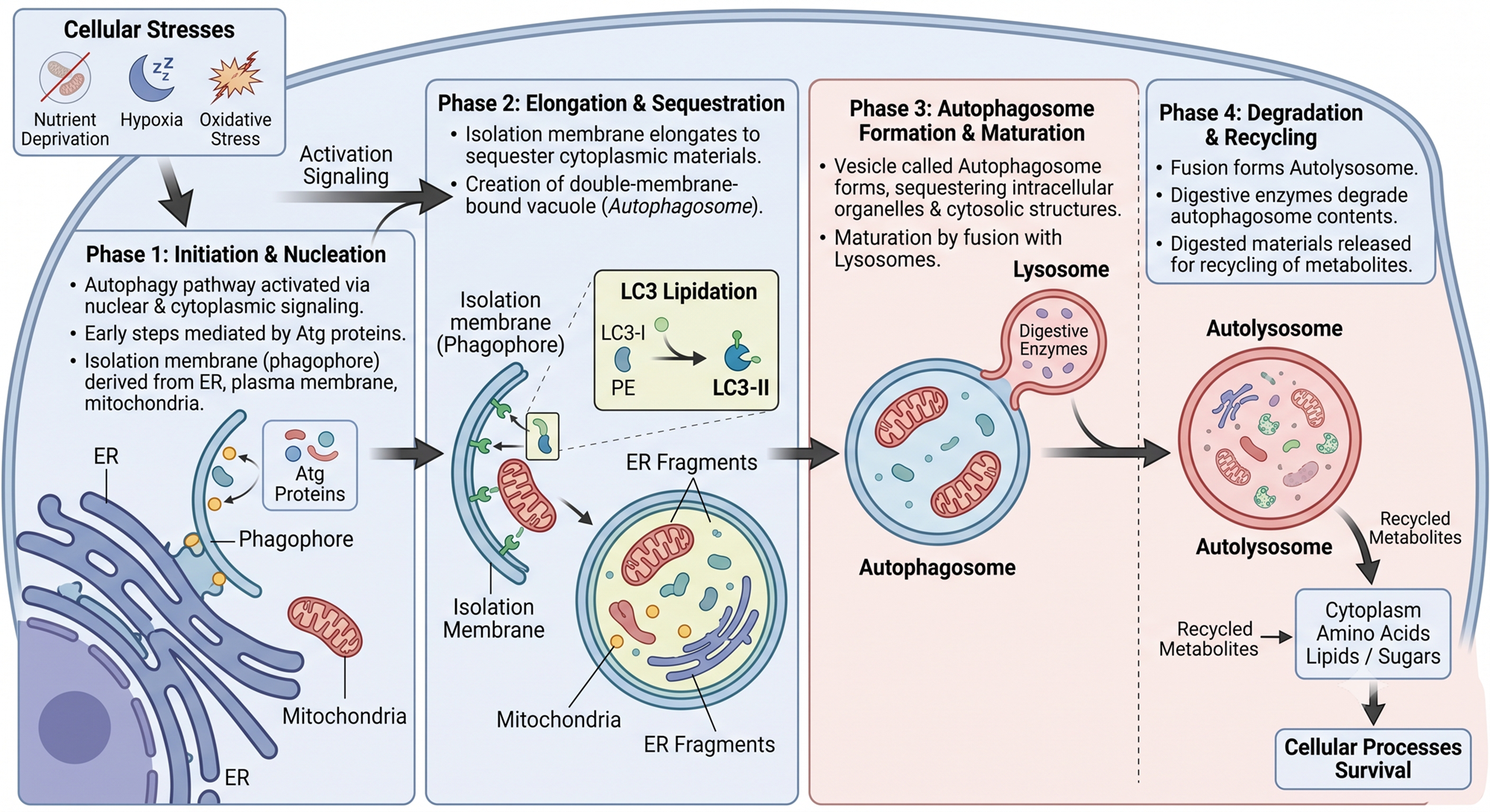
Autophagy is the cell's own cleaning and recycling system. Worn-out organelles, damaged proteins and other cytoplasmic material are wrapped in a membrane, carried to the lysosome, broken down, and the building blocks are used again. The name comes from Greek: auto (self) and phagy (eating).
Yoshinori Ohsumi received the 2016 Nobel Prize for working out how this system operates.
When does a cell switch on autophagy?
| Situation | What autophagy achieves |
|---|---|
| Starvation or nutrient shortage | Breaks down the cell's own parts to supply amino acids and energy |
| Loss of growth factors | Keeps the cell alive on fewer resources |
| Hypoxia, oxidative stress | Removes damaged organelles and membranes |
| Aging, exercise | Routine housekeeping: old parts out, room for new ones |
| Infection | Traps and destroys microbes inside the cell |
Autophagy is first of all a survival mechanism, and it is conserved from yeast to humans.
Types of autophagy
- Macroautophagy – the main type, and what "autophagy" usually means. A double-membrane vesicle (autophagosome) forms around the cargo.
- Microautophagy – the lysosomal membrane folds inward and takes in a small piece of cytoplasm directly.
- Chaperone-mediated autophagy – chaperone proteins recognise specific proteins and pass them across the lysosomal membrane; no vesicle forms.
The steps
Nutrient shortage → mTOR off, AMPK on
The initiation complex of Atg proteins (ULK1) is activated
A phagophore (isolation membrane) appears near the ER (Beclin-1 complex)
LC3 is joined to PE; the membrane grows around the cargo
The membrane seals → a double-membrane vesicle
Joins a lysosome → autophagolysosome
Lysosomal hydrolases digest the cargo; amino acids and fatty acids are reused
Points to note
- The phagophore is a cup-shaped starting membrane. Its main source is thought to be the ER, with possible contributions from mitochondria and the plasma membrane.
- The autophagosome is recognised by its double membrane, which is how it is identified on electron microscopy.
- In the autophagolysosome, both the inner membrane and the trapped contents are digested.
Molecular mechanism – what to remember for exams
Atg genes More than 30 autophagy-related genes (Atg) are known. Their protein products assemble in a fixed order to build the autophagosome.
Control: mTOR and AMPK
- When nutrients are plentiful, mTOR is active and suppresses autophagy.
- In starvation, mTOR switches off and falling energy activates AMPK, which starts autophagy.
LC3 – the marker of autophagy
- Two ubiquitin-like conjugation systems attach the lipid phosphatidylethanolamine (PE) to the protein LC3.
- Plain LC3 = LC3-I (in the cytosol). PE-linked LC3 = LC3-II (on the autophagosome membrane).
- LC3-II rises when autophagy is active, so it is used as a laboratory marker.
Selective autophagy Autophagy does not swallow cytoplasm blindly. Lipidated LC3, working with adaptor proteins such as p62, picks out particular cargo:
| Name | What is removed |
|---|---|
| Mitophagy | Damaged mitochondria |
| Aggrephagy | Protein aggregates |
| Xenophagy | Intracellular microbes |
| ER-phagy | Excess or damaged ER |
Autophagy and cell death
- In most settings autophagy protects the cell by recycling metabolites and clearing debris.
- It is prominent in atrophy, where cells shrink under severe nutrient shortage, and autophagic vacuoles are easy to find.
- It also handles normal turnover of ER, mitochondria and lysosomes.
- If the stress is too great, cell death pathways can be switched on: apoptosis, necroptosis and ferroptosis.
- Whether the cell dies from autophagy itself or from the stress that triggered it is still unsettled.
Autophagy in disease
| Disease | Role of autophagy | Point to remember |
|---|---|---|
| Cancer | Double-edged: can restrain tumours and can also help tumour cells survive stress | Both protective and promoting |
| Alzheimer disease | Autophagosome maturation is impaired | Protein aggregates build up |
| Huntington disease | Mutant huntingtin disturbs autophagy | Neurodegeneration |
| Tuberculosis and other infections | Mycobacteria, Shigella and HSV-1 are destroyed by autophagy | Loss of Atg5 in macrophages raises TB susceptibility |
| Crohn disease, ulcerative colitis | Linked to SNPs in ATG16L1 | Genetic risk for IBD; mechanism unknown |
In infection there is a second benefit: digested microbial proteins become available for antigen presentation.
Quick revision
- Autophagy = lysosomal digestion and recycling of the cell's own components
- Main trigger: nutrient deprivation
- Main membrane source: ER
- Autophagosome: double-membrane vesicle
- Marker: LC3-II (PE-lipidated LC3)
- mTOR suppresses, AMPK induces
- IBD: ATG16L1; TB: Atg5; Alzheimer: impaired maturation
Test yourself – 10 MCQs
Which stimulus is the classic trigger of autophagy?
The isolation membrane (phagophore) is believed to arise mainly from the:
Which structure is bounded by a double membrane?
The most useful marker for identifying cells undergoing autophagy is:
Elongation and closure of the autophagosome membrane require:
Which kinase suppresses autophagy when nutrients are abundant?
Crohn disease and ulcerative colitis have been linked to polymorphisms in which gene?
Deleting Atg5 specifically in macrophages increases susceptibility to:
The autophagy defect characteristic of Alzheimer disease is:
Which statement about autophagy is FALSE?
These notes are written in my own words for learning and teaching. Further reading: Robbins & Cotran Pathologic Basis of Disease, Chapter 2 (Cell Injury, Cell Death, and Adaptations).