Amyloidosis *Pathology notes: definition, pathogenesis, classification, morphology, clinical features અને diagnosis*
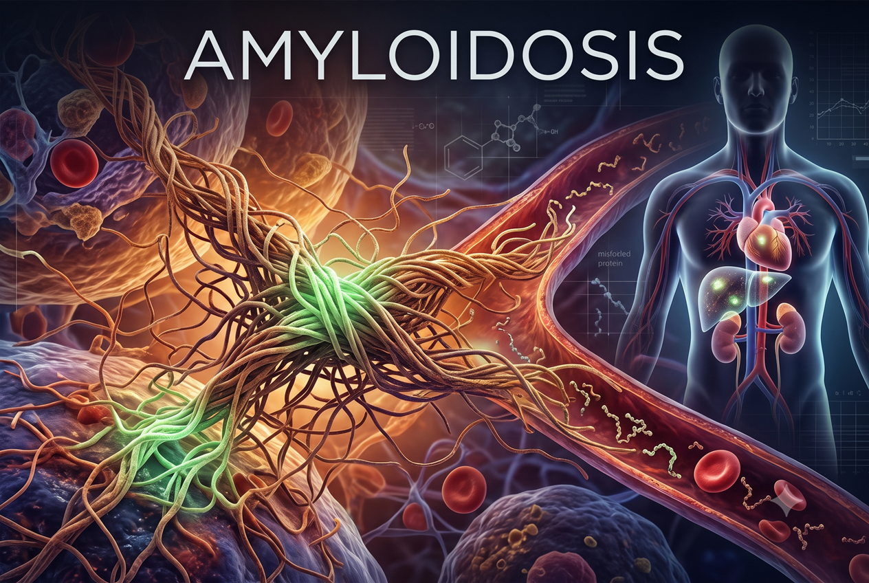
Pathology notes: definition, pathogenesis, classification, morphology, clinical features અને diagnosis
Amyloidosis pathology નો એવો topic છે જેમાં protein chemistry, histology અને clinical medicine એકસાથે આવે છે. આ notes માં amyloid કેવી રીતે બને છે, તેના types, organ-wise morphology, clinical features અને diagnosis step by step સમજાવ્યા છે. અંતે exam માટે quick points પણ આપ્યા છે.
1. Amyloidosis એટલે શું?
- Amyloidosis એવી disorders નું group છે જેમાં misfolded proteins aggregate થઈને fibrils બનાવે છે અને extracellular space માં deposit થાય છે.
- Deposits ધીમે ધીમે વધે છે, આસપાસના cells પર દબાણ લાવે છે, tissue damage કરે છે અને organ function ઘટાડે છે.
- Amyloidosis એક disease નથી. તે diseases નું group છે, જેમાં common વાત એ છે કે બધામાં એકસરખા દેખાતા proteins deposit થાય છે.
2. Fibril Formation ની પ્રક્રિયા
Soluble protein → Oligomers → મોટા aggregates → Insoluble fibrils → Tissue deposition
 Soluble protein misfold થઈને oligomers, aggregates અને છેવટે insoluble fibrils બનાવે છે.
- Normal folded configuration માં આ proteins soluble હોય છે.
- Misfold થયા પછી તે નાના groups (oligomers) બનાવે છે, પછી મોટા aggregates, અને છેવટે insoluble fibrils.
3. Deposit નું Composition અને "Amyloid" નામ
- આશરે 95%: fibril proteins
- આશરે 5%: serum amyloid P (SAP) અને બીજા glycoproteins
- Deposits સાથે proteoglycans અને glycosaminoglycans (heparan sulfate, dermatan sulfate) bind થાય છે.
- તેમાં charged sugar groups હોવાથી staining starch (amylose) જેવી લાગી, એટલે "amyloid" નામ પડ્યું. Deposit ને starch સાથે કોઈ સંબંધ નથી.
4. Physical Nature (Ultrastructure)
- Electron microscopy: continuous, nonbranching fibrils, diameter 8–10 nm
- દરેક fibril માં β-pleated sheet structure ધરાવતા protofilaments ના stacks હોય છે.
- આ cross-β-pleated conformation ને કારણે Congo red staining અને apple-green birefringence મળે છે.
- Amyloid નો type ગમે તે હોય, physical organization uniform હોય છે.
 Electron microscopy: 8–10 nm diameter ધરાવતા nonbranching fibrils.
 Fibril નું β-pleated sheet conformation.
5. Chemical Nature: Biochemical Diversity
- 40 થી વધુ જુદા જુદા proteins amyloid બનાવી શકે છે.
- 3 major forms (AL, ATTR, AA) અને કેટલાક minor forms છે.
(a) AL: Amyloid Light Chain
- Complete immunoglobulin light chains, તેમના amino-terminal fragments, અથવા બંને
- મોટા ભાગે λ (lambda) chains; κ (kappa) chains પણ amyloid બનાવી શકે
- Monoclonal plasma cells દ્વારા secrete થતી free Ig light chains માંથી બને છે
- Multiple myeloma જેવા plasma-cell tumors સાથે associated
(b) ATTR: Transthyretin Amyloid
- TTR એક plasma protein છે જે thyroxine અને retinol ને transport કરે છે
- ATTRwt (wild-type): normal TTR aggregate થાય છે; મુખ્યત્વે heart; સામાન્ય રીતે 70 વર્ષથી મોટા males
- ATTRv (variant): heart, peripheral nerves અને બીજા tissues
(c) AA: Amyloid-Associated
- Liver માં બનતા precursor SAA (serum amyloid-associated protein) ના proteolysis થી બને છે
- SAA blood માં HDL સાથે bound થઈને circulate થાય છે
- Inflammation માં acute-phase response દરમિયાન SAA વધે છે, એટલે AA ને chronic inflammation સાથે સંબંધ છે
Summary Table
| Type | Precursor / Source | Typical Setting |
|---|---|---|
| AL | Ig light chains (κ/λ) | Monoclonal plasma-cell disorders |
| ATTRwt | Wild-type transthyretin | Age-associated cardiac deposition |
| ATTRv | Variant transthyretin | Inherited cardiac / nerve disease |
| AA | SAA (liver) | Chronic inflammatory states |
Frequency: Systemic amyloidosis નું સૌથી common form AL છે, ત્યારબાદ ATTRwt. Population aging સાથે ATTRwt નું prevalence વધી રહ્યું છે.
(d) બીજા Important Amyloid Proteins
- β-amyloid (Aβ): Alzheimer disease ની cerebral plaques નો core, અને cerebral blood vessel walls માં deposit. મોટા transmembrane glycoprotein APP (amyloid precursor protein) માંથી બને છે.
- β2-microglobulin (Aβ2m): MHC class I નો component. Long-term hemodialysis patients માં joints અને soft tissues માં deposit થાય છે.
- Prion protein: કેટલાક CNS prion diseases માં misfolded prion proteins extracellular aggregates બનાવે છે, જેમાં amyloid જેવી staining characteristics હોય છે.
6. Pathogenesis
Quality control failure:
- Normally, misfolded proteins intracellularly proteasomes માં અથવા extracellularly macrophages દ્વારા degrade થાય છે.
- Amyloidosis માં આ quality-control mechanisms fail થાય છે, એટલે misfolded protein cells ની બહાર accumulate થાય છે.
Amyloid બનાવતા proteins ની 2 categories:
- Normal proteins જેમાં misfold થવાની inherent tendency હોય. વધુ માત્રામાં બને અથવા degradation impaired હોય ત્યારે amyloid બનાવે છે (દા.ત. AL, AA, ATTRwt).
- Variant (mutant) proteins જે misfolding માટે prone હોય (દા.ત. ATTRv).
Localized vs Systemic:
- Localized: protein ની production site પર જ deposit
- Systemic: production site થી દૂરના organs માં deposit
Organ dysfunction કેમ થાય છે?
- મુખ્યત્વે deposits ને કારણે થતા distorted tissue architecture થી
- વધુમાં, કેટલાક amyloidogenic proteins ROS production વધારે છે અને calcium homeostasis બગાડે છે
Classification: Deposit થયેલા protein ના biochemical nature પરથી નામ અપાય છે (AL, ATTR, AA…).
7. Classification in Detail
AL Amyloidosis (પહેલાં "Primary Amyloidosis")
- સામાન્ય રીતે systemic; સૌથી common form (USA માં દર વર્ષે આશરે 2000–3000 new cases)
- Cause: plasma cells નું clonal proliferation, જે amyloid-prone light chain બનાવે છે
- Multiple myeloma ના 5–15% patients માં systemic amyloidosis થાય છે
- Myeloma માં:
- Single Ig વધુ બને છે, જે serum electrophoresis પર M-protein spike આપે છે
- Free, unpaired κ અથવા λ light chains = Bence-Jones protein
- નાનું size હોવાથી urine માં excrete થાય છે
- મોટા ભાગના myeloma patients ને free light chains હોવા છતાં amyloidosis થતું નથી. Light chain નું amino acid sequence તેનું amyloidogenic potential નક્કી કરે છે.
- AL ધરાવતા મોટા ભાગના patients ને overt myeloma હોતો નથી, પણ:
- Serum/urine માં monoclonal Ig અથવા free light chains મળે છે
- Bone marrow માં plasma cells થોડા વધેલા હોય છે
- એટલે underlying monoclonal gammopathy હોય છે, જેમાં tumor mass કરતાં abnormal protein production મુખ્ય સમસ્યા છે
ATTR (Transthyretin) Amyloidosis
- Genetic mutation અથવા protein aging ને કારણે TTR misfolding થાય છે, જેમાંથી insoluble fibrils બને છે
- ATTRwt: heart અને connective tissues (દા.ત. ligaments); 70 વર્ષથી મોટા males; બીજું સૌથી common systemic form
- ATTRv: autosomal dominant
- Familial amyloid polyneuropathy: peripheral અને autonomic nerves
- Familial cardiomyopathy (ATTRv-CM): heart predominant
- USA માં African-American population ના આશરે 4% લોકો એક cardiac TTR variant carry કરે છે (origin: West Africa). Homozygous અને heterozygous બંનેમાં restrictive cardiomyopathy થઈ શકે.
- Penetrance variable છે, એટલે કોઈને severe disease થાય અને કોઈને કંઈ ન થાય
AA Amyloidosis (પહેલાં "Secondary / Reactive Amyloidosis")
- Systemic, AA protein ના deposits
- પહેલાંના causes: tuberculosis, bronchiectasis, chronic osteomyelitis (antibiotics આવ્યા પછી ઘટ્યા)
- હાલના common causes:
- Rheumatoid arthritis: સૌથી common association; RA ના આશરે 3% patients માં amyloidosis થાય છે, અને તેમાંથી અડધામાં clinically significant
- Ankylosing spondylitis
- Inflammatory bowel disease (Crohn disease, ulcerative colitis)
- Heroin નું subcutaneous injection ("skin-popping"), જેમાં chronic skin infections થાય છે
- Tumors: renal cell carcinoma, Hodgkin lymphoma
- Mechanism:
- Inflammation માં IL-6, IL-1 liver માં SAA synthesis વધારે છે
- Long-standing inflammation હોય તો SAA સતત ઊંચું રહે છે
- ફક્ત SAA વધવું પૂરતું નથી. Macrophage enzymes SAA ને normally degrade કરે છે. Amyloidosis થવા માટે કદાચ enzyme defect (incomplete breakdown) અથવા structurally abnormal SAA (degradation-resistant) જરૂરી છે.
- Underlying diseases ની સારવાર સુધરી હોવાથી prevalence ઘટી રહ્યું છે
8. Other Types of Amyloidosis
Heredofamilial Amyloidosis: Familial Mediterranean Fever (FMF)
- Autosomal recessive; સૌથી common અને best-studied familial form
- Autoinflammatory syndrome જેમાં IL-1 વધુ બને છે
- Fever ના attacks સાથે serosal surfaces (peritoneum, pleura, synovium) નું inflammation
- Gene pyrin encode કરે છે, જે inflammasome દ્વારા inflammation regulate કરે છે
- Armenian, Sephardic Jewish અને Arabic origin ના લોકોમાં વધુ જોવા મળે છે
- Amyloid type: AA (IL-1 → SAA વધે)
Hemodialysis-Associated Amyloidosis
- Protein: β2-microglobulin
- Kidney disease માં serum માં તેનું level ઊંચું હોય છે, અને જૂની dialysis membranes તેને filter કરી શકતી ન હતી
- Joints, muscle, tendons, ligaments માં deposits
- Classic presentation: carpal tunnel syndrome
- નવા dialysis filters આવ્યા પછી incidence ઘણું ઘટ્યું છે
Localized Amyloidosis
- એક જ organ અથવા tissue સુધી limited
- Nodular masses અથવા ફક્ત microscopic deposits
- Common sites: lung, larynx, skin, urinary bladder, tongue, periorbital region
- Periphery માં lymphocytes અને plasma cells નું infiltrate હોય છે; ઘણી વાર localized AL (plasma cell-derived)
 Localized amyloidosis: grossly દેખાતા nodular deposits.
Endocrine Amyloid
- Sites: medullary thyroid carcinoma, pancreatic islet tumor, pheochromocytoma, type 2 DM માં islets of Langerhans
- Source: polypeptide hormones (દા.ત. calcitonin, medullary carcinoma માં) અથવા unique proteins (દા.ત. islet amyloid polypeptide / amylin)
- Medullary carcinoma of thyroid માં amyloid ની હાજરી diagnostic clue છે
9. Amyloid Precursors: Quick Reference
Systemic:
- AL: Ig light chain (primary / myeloma-associated); AH: Ig heavy chain (rare variant)
- AA: SAA (secondary / reactive, FMF)
- Aβ2M: β2-microglobulin (hemodialysis; synovium, bone)
- ATTR: transthyretin (familial + age-related; heart, nerves, soft tissue)
- Rare familial forms: apolipoprotein AI/AII, gelsolin, fibrinogen Aα, lysozyme, LECT2 (મુખ્યત્વે renal / hepatic involvement)
Localized:
- Aβ: Alzheimer disease, Down syndrome (CNS)
- ACys: cystatin C (cerebral amyloid angiopathy)
- APrP: prion protein (spongiform encephalopathies)
- AIAPP: amylin (type 2 DM, pancreas)
- ACal: calcitonin (medullary carcinoma of thyroid)
- AANF: atrial natriuretic factor (cardiac atria, atrial fibrillation)
- APro: prolactin (pituitary)
10. Morphology
Organ Distribution
- AA (chronic inflammation): kidneys, liver, spleen, lymph nodes, adrenals, thyroid
- AL (plasma-cell associated): વધુ વાર heart, GI tract, respiratory tract, peripheral nerves, skin, tongue
- Hereditary: variable; FMF માં widespread (kidney, vessels, spleen, respiratory tract)
- Distribution હંમેશા diagnostic રીતે distinctive હોતું નથી
Gross
- મોટા deposits હોય તો organ enlarged, gray, waxy, firm બને છે
Microscopy
- H&E: amorphous, eosinophilic, hyaline, extracellular material
- Deposition હંમેશા extracellular હોય છે, cells ની વચ્ચે, ઘણી વાર basement membrane ની નજીક શરૂ થાય છે
- વધતા deposits cells ને ઘેરીને destroy કરે છે
- AL માં perivascular અને vascular deposits common છે
- Collagen અને fibrin થી અલગ ઓળખવા special stains જરૂરી છે
 H&E: cells ની વચ્ચે amorphous, eosinophilic, hyaline amyloid deposits.
 Basement membrane પાસે શરૂ થતું extracellular deposition, જે વધતાં cells destroy કરે છે.
Congo Red Staining
- Ordinary light: pink/red
- Polarized light: apple-green birefringence, જે બધા amyloid types માં common છે અને cross-β-pleated configuration ને કારણે થાય છે
- EM: thin, nonbranching, nonoriented fibrils
- Typing: immunohistochemistry (AA, AL, TTR) અને mass spectrometry (બધા types)
 Congo red, ordinary light: amyloid deposits pink-red દેખાય છે.
 Congo red, polarized light: characteristic apple-green birefringence.
Kidney: સૌથી common અને સૌથી serious
- Gross: normal size, અથવા advanced cases માં shrunken (vascular narrowing થી ischemia)
- મુખ્યત્વે glomeruli; સાથે interstitium, arteries, arterioles
- Progression: mesangial thickening → basement membrane નું uneven widening → capillary narrowing → capillary lumens obliterate → glomerulus amyloid ના confluent masses અથવા broad ribbons થી ભરાઈ જાય
 Glomerular amyloid: early mesangial deposits થી advanced confluent amyloid અને obliterated lumen સુધી.
Spleen
- Inapparent થી marked splenomegaly (800 g સુધી)
- Sago spleen: deposits splenic follicles માં; tapioca-like granules
- Lardaceous spleen: red pulp ના sinus walls અને connective tissue framework માં; deposits ભેગા થઈને map-like areas બનાવે
 Sago spleen (follicular deposits) અને lardaceous spleen (red pulp, map-like areas).
Liver
- Hepatomegaly થઈ શકે
- Deposits પહેલાં space of Disse માં, પછી hepatocytes અને sinusoids પર encroach
- Pressure atrophy થી hepatocytes ગાયબ થાય છે, અને parenchyma ના મોટા areas replace થાય છે
- Vascular involvement frequent છે
- Extensive involvement હોવા છતાં liver function સામાન્ય રીતે preserved રહે છે
 Liver: space of Disse થી શરૂ થઈને hepatocyte loss સુધી.
Heart
- Systemic amyloidosis ના કોઈ પણ form માં; senile systemic (ATTRwt) માં heart major organ છે
- Gross: enlarged અને firm, અથવા ઘણી વાર normal દેખાય
- Deposits: subendocardial અને myocardial fibers ની વચ્ચે, જેનાથી pressure atrophy થાય
- Subendocardial deposits conduction system damage કરે છે, એટલે ECG abnormalities અને arrhythmias થાય
 Heart: myocardial fibers ની વચ્ચે amyloid deposits.
Other Organs
- Tongue: macroglossia ("tumor-forming amyloid of tongue")
- Respiratory tract: larynx થી નાની bronchioles સુધી
- Brain: Alzheimer disease માં plaques અને vessels
- Peripheral / autonomic nerves: familial amyloidotic neuropathies
- Hemodialysis: wrist નું carpal ligament (median nerve compression), joints
11. Clinical Features
- Amyloidosis incidental histologic finding પણ હોઈ શકે, અને fatal disease પણ.
- Symptoms deposits ની magnitude અને involved organs પર આધાર રાખે છે.
Early (nonspecific): weakness, weight loss, lightheadedness, syncope
Renal:
- Proteinuria → nephrotic syndrome
- Glomeruli નું progressive obliteration → kidney failure → uremia
- Kidney failure death નું common cause છે
Cardiac:
- Insidious congestive heart failure
- સૌથી serious: conduction disturbances અને arrhythmias (fatal હોઈ શકે)
- Restrictive cardiomyopathy, જે chronic constrictive pericarditis જેવી લાગી શકે
GI:
- Macroglossia, જેનાથી speech અને swallowing માં તકલીફ થાય
- Malabsorption, diarrhea, digestive disturbances
Vascular / Bleeding:
- Vascular fragility, જેમાં નાના trauma થી પણ bleeding (ક્યારેક massive) થાય
- AL amyloid factor X ને bind કરીને inactivate કરે, જેનાથી life-threatening bleeding થઈ શકે
AL amyloidosis ના classic clinical signs:
- Macroglossia
- Periorbital ecchymoses ("raccoon eyes")
- Fingernail dystrophy
 AL amyloidosis ના clinical signs.
12. Diagnosis
Tissue Diagnosis (Congo red)
- Abdominal fat aspirate: noninvasive; specific પણ sensitivity low
- Rectal અથવા gingival biopsy (systemic amyloidosis ની શંકા હોય ત્યારે)
- Kidney biopsy (renal manifestations હોય ત્યારે)
Diagnostic Approach (Stepwise)
- Clinical suspicion: unexplained nephropathy, cardiomyopathy, neuropathy, enteropathy, arthropathy, macroglossia
- Noninvasive biopsy (abdominal fat)
- Positive → typing
- Negative → affected organ ની invasive biopsy; તે પણ negative હોય તો further work-up નહીં
- Typing: mass spectrometry અથવા IHC
- κ / λ light chain → monoclonal protein અને bone marrow plasma cells શોધો → AL (heart, kidney, liver, autonomic involvement અને factor X deficiency માટે screen કરો)
- Amyloid A → underlying chronic inflammatory disease શોધો → AA
- Transthyretin → mutant હોય તો ATTRv (relatives ને screen કરો); wild-type હોય તો ATTRwt (સામાન્ય રીતે >65 વર્ષના males, cardiac)
- Negative → rare familial types (ApoAI, ApoAII, fibrinogen, lysozyme, gelsolin)
 Clinical suspicion → biopsy → typing → underlying cause.
AL માટે Laboratory Work-up
- Serum અને urine protein electrophoresis + immunofixation
- Serum protein electrophoresis ઘણી વાર normal હોય છે, એટલે immunofixation વધુ sensitive છે (દા.ત. IgGκ monoclonal protein)
- Bone marrow:
- Overt myeloma વગર પણ monoclonal plasmacytosis
- CD138 (syndecan) IHC થી plasma cells ઓળખાય
- κ અને λ mRNA in situ hybridization થી clonality બતાવી શકાય
 AL work-up: serum immunofixation માં monoclonal band; bone marrow માં clonal plasma cells.
Imaging
- Radiolabeled SAP scintigraphy: rapid અને specific. Amyloid deposits સાથે bind થાય છે, extent બતાવે છે અને treatment follow-up માં ઉપયોગી છે.
13. Prognosis અને નવી Therapy
- Generalized amyloidosis નું prognosis poor છે.
- AL: diagnosis પછી median survival આશરે 2 વર્ષ; myeloma-associated AL માં તેનાથી પણ ખરાબ.
- Reactive (AA): થોડું સારું prognosis; underlying disease ના control પર આધાર. Amyloid નું resorption report થયું છે, પણ rare છે.
- નવી દિશાઓ:
- Plasma cell tumors માટે નવી, વધુ effective therapies
- Protein misfolding સુધારવી અને fibrillogenesis inhibit કરવી
Quick Exam Points
- Amyloid = extracellular, misfolded protein ના β-pleated sheet fibrils, 8–10 nm, nonbranching
- Composition: 95% fibril protein + 5% SAP / glycoproteins
- Congo red: pink-red in ordinary light, apple-green birefringence in polarized light
- Most common systemic form: AL, ત્યારબાદ ATTRwt
- AL: λ > κ; myeloma ના 5–15% માં; મોટા ભાગના AL patients ને overt myeloma નથી
- AA: SAA (liver, HDL-bound); IL-6, IL-1; સૌથી common cause rheumatoid arthritis
- ATTRwt: elderly males, heart; ATTRv: autosomal dominant (nerves / heart)
- FMF: autosomal recessive, pyrin, IL-1, AA amyloid
- Hemodialysis: β2-microglobulin, carpal tunnel syndrome
- Alzheimer: Aβ from APP
- Medullary thyroid carcinoma: calcitonin-derived amyloid, diagnostic clue
- Kidney: most common and most serious; nephrotic syndrome; kidney failure common cause of death
- Spleen: sago (follicles) vs lardaceous (red pulp)
- Liver: space of Disse; function preserved
- Heart: subendocardial deposits, arrhythmias, restrictive cardiomyopathy
- AL signs: macroglossia, periorbital purpura, nail dystrophy, factor X deficiency
- Fat aspirate: specific, low sensitivity; SPEP may be normal, so do immunofixation
- AL median survival ≈ 2 years